Interview | Dr. Habib Zaidi, from anatomical to molecular imaging technology
Dr. Habib Zaidi is Chief Physicist and Head of the Positron Emissions Tomography (PET) Instrumentation & Neuroimaging Laboratory at Geneva University Hospital. He is also a Professor of Medical Physics at the University Medical Center of Groningen (the Netherlands) and visiting Professor at École Nationale Supérieure d’Electronique et de ses Applications (ENSEA, France). Dr. Zaidi is actively involved in developing imaging solutions for cutting-edge interdisciplinary biomedical research and clinical diagnosis. His academic accomplishments in the area of quantitative PET imaging have been well recognized through many awards and distinctions most notably the prestigious 2010 Kuwait Prize of Applied Sciences (also known as the Middle Eastern Nobel Prize) awarded by the Kuwait Foundation for the Advancement of Sciences (KFAS), which he received for outstanding accomplishments in Biomedical Technology.
Dr. Zaidi speaks to Inspire Magazine about the latest advances in molecular imaging technologies, his contributions to the field which led him to receiving the 2010 Kuwait Prize of Applied Sciences, as well as the status of biomedical research in Algeria and the potential for collaborations between Algerian scientists and practitioners inside and outside of the country.
Inspire magazine: Dr. Habib Zaidi, many thanks for speaking to Inspire magazine. In simple terms, could you please explain to us what is your area of research and what led you to this specialisation?
 HZ: I graduated in Electrical Engineering from the University of Setif in Algeria and then was attracted to the fascinating world of medical physics and started my postgraduate program to end-up with a Ph.D. awarded by Geneva University followed by habilitation (Privat-docent) in the same field. My area of expertise is medical imaging physics and instrumentation and my lab is actively involved in developing imaging solutions for cutting-edge interdisciplinary biomedical research and clinical diagnosis. My research focuses on Positron Emission Tomography (PET), which is a medical imaging technique that allows doctors and researchers to investigate biochemical and biological processes in vivo, in living organisms, using radioactive tracers. This is particularly relevant in modern day medicine due to the high interest in molecular imaging-guided assessment of response to treatment and radiation therapy planning.
IM: How effective are currently used medical imaging techniques and what are their main limitations?
HZ: Traditional imaging methods such as plain film radiography, ultrasound and more recent techniques such as x-ray computed tomography (CT) and magnetic resonance imaging (MRI) are very useful to evaluate a patient’s anatomy with sub-millimeter spatial resolution, which allows doctors to detect any structural abnormalities and to evaluate the location and extent of disease. These methods also offer relatively fast scan times, and the resulting scans can be easily interpreted due to good tissue contrast especially when contrast media are administered to the patient.
Methods such as x-ray projection imaging, CT and MRI differentiate diseased from normal tissue by looking at the structural differences in tissues or differences in regional perfusion of the administered contrast media. However, in some instances where the normal perfusion patterns of the tissues have been disrupted due to prior surgery or radiation therapy, the resulting tissue damage or necrosis can show contrast patterns that mimic those associated with neoplasia, which complicates the interpretation of the scans and could lead to misdiagnosis. This is particularly crucial and presents a significant challenge when the imaging techniques are used to determine patients’ response to treatment, making it difficult to determine the exact anatomical extent of disease when planning conformal radiation treatment or for planning highly targeted therapeutic regimes.
In comparison to these anatomical imaging techniques, functional imaging methods including planar scintigraphy, single-photon emission computed tomography (SPECT), positron emission tomography (PET), and magnetic resonance spectroscopy (MRS) focus on the regional differences in the biochemical composition of tissues. In other words, these techniques examine the molecular level to differentiate diseased tissue from healthy tissue to give more precise results. This makes these detection techniques much more sensitive, in fact they are about 109 more sensitive compared to macroscopic medical imaging techniques such as CT and MRI.

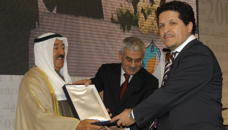
Dr. Habib Zaidi on the reception of his 2010 Kuwait Prize for Applied Sciences at the Kuwait Foundation for the Advancement of Sciences
IM: You have recently received the Kuwait Prize for Applied Sciences for outstanding contributions to Biomedical Technology. Many congratulations on this achievement. Could you please tell us about the essence of these contributions and how they improve on currently available imaging techniques?
HZ: In 2000, I initiated a basic research program and founded the PET Instrumentation and Neuroimaging laboratory (PinLab), which is part of the Geneva Neuroscience Center. In a relatively short time, the group has assumed a leading role in Switzerland and become internationally recognized for excellence in medical imaging research with PET and hybrid imaging (PET-CT and PET-MR) being a focus for its activities. Our group gained international recognition for contributions to the development and analysis of new image correction and reconstruction techniques for improved quantification of PET images as well as development and better understanding of PET Monte Carlo modeling tools. The contributions relate to the development of image reconstruction techniques, modeling/simulation tools and accurate data correction techniques for PET, as well as the assessment of new possible designs of PET detection modules and development of novel hybrid imaging technologies including PET-MR. This aims to further improve the accuracy of these imaging techniques and facilitate the interpretation and analysis of multimodality imaging data.
IM: So what is the ultimate potential that you would like to achieve with this collection of work?
HZ: We are convinced that molecular imaging will be the way to go for personalized medicine and the hope is to contribute to the improvement of current and development of novel technologies and methodologies to make this happen. We hope that some of our research activities will result in improving diagnosis and treatment and minimizing patients’ suffering.
")
Examples of molecular imaging techniques (left to right: PET-CT, PET-MR and PET)
IM: With this ultimate potential in mind, how do you see the future of medical imaging?
HZ: The clinical role of multimodality imaging encompasses a wide variety of applications and is now performed routinely with commercially available radiopharmaceuticals to answer important clinical questions including those in oncology, cardiology, neurology and psychiatry. Nowadays, a plethora of novel tracers are used routinely for assessing tumor metabolism and other biological and physiological parameters associated with many diseases that have clearly demonstrated the enormous potential of emerging hybrid technologies in the field of molecular imaging. In the near future, imaging will be used substantially in drug development and will play a significant role in personalized medicine.
IM: In general, how would you describe the status of biomedical research in Algeria and how can it be improved?
HZ: I was able to form ongoing collaborations with Algerian colleagues in the field of medical imaging through participating in various conferences and symposia in Algeria. From these experiences, it is clear that Algeria does not lack high profile scientists. Nonetheless, the research efforts carried in Algeria are far away from what we can expect with the quality of scientists in charge of carrying out the research programs in the country.
It is difficult to advise on improving the research status in Algeria without knowing in detail the reasons why it is lagging behind, but in essence there seems to be a huge bureaucratic power and lack of motivation. There is a strong need to train fresh and motivated scientists and give them the chance to establish themselves by giving them access to tools and instruments needed to excel in their research.
IM: What are the potentials and challenges for nurturing collaborations between specialist biomedical researchers inside and outside of Algeria?
 HZ: The opportunities are there and there are many good initiatives (summer & winter schools, conferences and symposia, official links through some non-governmental societies e.g. ACA). However, to be honest, most of the collaborations I had required a lot of investment in terms of time and resources to provide technical expertise and training, but the return on investment remains to be seen. The main challenge is the lack of a “political will” to make this happen and the lack of motivation of Algerian scientists given the conditions there.
IM: Are you aware of the imaging techniques currently used in the Algerian healthcare system and how advanced these are?
HZ: Yes, all conventional imaging technologies are available especially in the private sector. However, not all Algerians can afford the high cost of medical examinations such as CT and MRI and there is a lack of support for this category of patients to get scanned in public facilities. Advanced molecular imaging technologies are still not introduced, despite the fact that we were invited as early as 2006 to advice the Algerian government on how to establish such programs in the country.
The state-of-the-art technological developments in medical imaging cannot be viewed as a uniform reality all over the world. If we take the example of multimodality medical imaging, we see that despite the commercial introduction of dual-modality imaging systems and its widespread acceptance in clinical settings in various parts of the world, the healthcare system in our country is lagging behind. So, while cross-training requirements and guidelines for both the technologists who operate the combined units and the physicians (radiologists and nuclear medicine physicians) who interpret the images are being debated in developed nations, the clinical relevance of this promising technology is still being questioned in Algeria and many other developing countries.
It will probably take a while before clinicians in our country will be prepared to adjust the way they are used to practice medicine to take full advantage of novel state-of-the-art technologies available today. Moreover, despite the favorable economic situation and the availability of financial resources in Algeria, it might take a while before politicians and decision makers realize the potential and cost-effectiveness of these novel technologies.
IM: How do you think the standard of biomedical imaging in the Algerian healthcare system can be improved in terms of existing or potential collaborations between researchers and practitioners inside and outside of Algeria? For example, what practical steps could be taken to leverage these collaborations for the benefit of the healthcare system?
HZ: I am under the impression that, to date, there is a lack of knowledge of the currently available imaging equipment throughout the country and the potential of existing research groups. I believe it is very important at this stage to carry out an audit at the national level to have a clear picture of the current situation and then start from there to guarantee that the main university hospitals throughout the country are well equipped with the needed medical imaging technologies and appoint qualified scientists to operate them and exploit them to their full potential. These scientists can then take the lead at the national level and maintain bridges with their counterparts in the developed world.
IM: Your area of research makes use of advanced technological equipment. What would you advise biomedical researchers in Algeria who may not necessarily have access to such technology and who wish to conduct cutting edge research?
HZ: I have a strong opinion on this issue. It is unfortunate to note that most developing countries still rely on developed countries to provide education and training programs while parts of the needed equipment is provided through international donations of used equipment. These problems of availability and needed implementation of strategies for cancer diagnosis and treatment are common in developing countries. From my own experience, it appears that the optimization of current resources is more important than obtaining additional equipment and resources. Adequate education, training and application of standard diagnostic and therapeutic approaches and techniques should improve healthcare delivery.
The past century was the century of “big hit science” for medical imaging where major discoveries and inventions were brought to the world by brilliant scientists that revolutionized the practice of medicine. Medical imaging science is not an undemanding and easy profession, it should never be considered as a ‘stomach job’ used to earn one’s living, but instead as a passion that should be given the place it deserves in our lives. The words of late Prof. Abdus Salam (1979 Nobel Laureate in Physics) come to mind: “Scientists are very happy people because their job is also their hobby”.
IM: Many Thanks again for taking the time to speak to Inspire magazine and all the best with your future endeavours.
HZ: Thank you very much for giving me this opportunity.
Romaissa Asme Boussahel author
Dr. Romaissa Asme Boussahel obtained her PhD from the School of Pharmacy, University of Nottingham, her work focuses on drug delivery, tissue engineering and regenerative medicine with a particular focus on growth factor delivery formulations and bone regeneration. She is Co-founder and Associate Editor of Inspire Magazine.
3 Comments so far
Lotf ZAIDIPosted on 8:09 pm - Nov 29, 2012
Congratulations for all these acheavements.You are our pride, more success in the future.
Hacene ZAIDIPosted on 6:12 pm - Dec 7, 2012
Thank you HABIB and congrtulations for all your contributions.
SamiaPosted on 12:35 am - Feb 18, 2013
Nice interview, an excellent scientist but also a respectable human being …
About the Author